India charts a new atlas of the brainstem
2026-07-13
A map, not a theory, is quietly resetting expectations about the human brain. An Indian team has assembled a high‑resolution brainstem atlas that dissects this compact structure into dozens of nuclei and fiber tracts with a precision most clinicians have never had on screen.
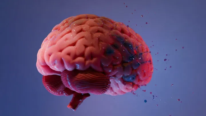
What sounds like cartography is really a bet on anatomy over guesswork. Using ultra‑fine magnetic resonance imaging and painstaking histological sectioning, researchers aligned structural detail down to sub‑millimeter scales, so that regions controlling respiration, cardiovascular reflexes, and sleep‑wake regulation can be distinguished rather than approximated in a blurry column of tissue.
The bolder claim is that this atlas will quietly change clinical routine before it rewrites textbooks. Neurosurgeons now gain a reference frame for deep brain stimulation targets near the periaqueductal gray or locus coeruleus; neuroradiologists can anchor signal changes in disorders such as multiple system atrophy to specific nuclei instead of vague zones; and basic scientists can plug the dataset into connectomics and tractography pipelines to test how ascending and descending pathways actually traverse this crowded junction of the nervous system.
Loading...

