PET/MRI Finds Hidden Seizure Sources
2026-09-12
PET/MRI can expose a seizure source when structural MRI leaves no convincing mark. That matters. In a large pediatric cohort with hard-to-classify epilepsy, the hybrid scan localized candidate epileptogenic zones even after negative or equivocal MRI findings, giving presurgical teams evidence that conventional anatomy had withheld.
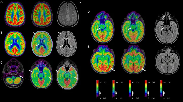
This is not merely a sharper picture. PET maps radiotracer distribution, reflecting cerebral glucose metabolism, while MRI charts anatomy and structural abnormalities. Together, the modalities work like a debugger tracing a rogue background process: metabolic disruption can flag where neuronal networks may be misfiring, then anatomical detail helps surgeons judge whether that signal belongs to tissue they can approach. The distinction is clinical, not cosmetic. Children with unresolved seizures may otherwise face longer monitoring, uncertain operative planning, or no surgical route at all.
The larger implication is hard to ignore. Earlier hybrid imaging could narrow invasive electrode placement and reduce missed lesions during presurgical evaluation. The remaining test is stern: better localization must yield safer resections and fewer seizures. At scale, fused metabolic and anatomical signals could turn the operating plan into a far more responsive decision engine.
Loading...

