MRI Maps Blood Flow Through Brain Layers
2026-09-17
This is a sharper lens. A new MRI measure from the Mark and Mary Stevens Neuroimaging and Informatics Institute at the Keck School of Medicine of USC noninvasively maps how cerebral blood flow aligns with cellular organization across layers of the living human brain. The scan reads depth. It links vascular signals to cytoarchitecture without surgery.
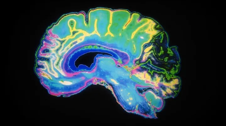
That pairing could change what a brain scan can say. It is less a still photograph than a systems monitor: MRI-derived cerebral blood flow becomes the live traffic signal, while cytoarchitecture supplies the street plan. The comparison is only a metaphor. In scientific terms, the approach joins layer-resolved hemodynamics with cellular organization, allowing researchers to examine whether tissue structure and perfusion correspond at distinct cortical depths. That matters because brain layers do not share identical cells, connections, or metabolic demands. Standard measures can blur those differences. This one tries to keep them visible.
For neuroscience, the appeal is not spectacle but resolution. By placing perfusion beside tissue architecture in the same living brain, the measure could expose mismatches hidden when either signal is examined alone, at a scale conventional summaries often flatten. No incision. No tissue sample. If validated across studies, this method could turn MRI into a layer-by-layer diagnostic engine, tracing where vascular support and cellular order begin to diverge before symptoms become obvious. The consequence could be a feedback system in which MRI does not merely show the brain, but flags the first places where its supply lines and cellular order fall out of step.
Loading...

